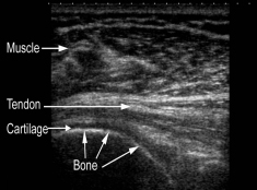
Ultrasound is safe and painless, and produces pictures of the inside of the body using sound waves. Ultrasound imaging, also called ultrasound scanning or sonography, involves the use of a small transducer (probe) and ultrasound gel placed directly on the skin. High-frequency sound waves are transmitted from the probe through the gel into the body. The transducer collects the sounds that bounce back and a computer then uses those sound waves to create an image.
Ultrasound examinations do not use ionizing radiation (as used in x-rays), thus there is no radiation exposure to the patient. Because ultrasound images are captured in real-time, they can show the structure and movement of the body’s internal organs, as well as blood flowing through blood vessels.
Ultrasound images are typically used to help diagnose:
- Tendinopathies, ligamentous sprains and muscle tears
- Inflammation or fluid (effusions) within the bursae and peripheral joints such as the shoulder, elbow, wrist, knee and ankle
- Early changes of rheumatoid arthritis.
- Nerve entrapments such as carpal tunnel syndrome.
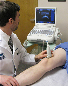
Charlie uses ultrasound both as a diagnostic instrument and for obtaining detailed images to assist him in administering guided injections.
To find out more or to book an appointment please contact us:
T: 01227 266439
E: info@joylaneclinic.co.uk